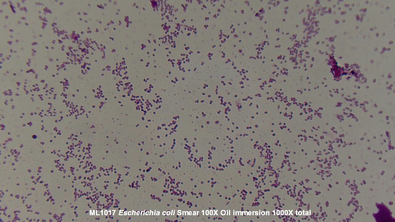
[最も選択された] e coli microscope 284026-E coli under microscope 1000x
Bacteria species E coli and S aureus under the microscope with different magnifications Bacteria are among the smallest, simplest and most ancient livingE coli can spread to the urinary tract in a variety of ways Common ways include Your urine will then be examined under a microscope for the presence of bacteria Urine cultureSUMMARY The shape of Escherichia coli is strikingly simple compared to those of higher eukaryotes In fact, the end result of E coli morphogenesis is a cylindrical tube with hemispherical caps It is argued that physical principles affect biological forms In this view, genes code for products that contribute to the production of suitable structures for physical factors to act upon

Figure 3 From Pathologicalstudy On Colibacillosis In Chickens And Detection Of Escherichia Coli By Pcr Semantic Scholar
E coli under microscope 1000x
E coli under microscope 1000x-Study E coli, the intestinal bacteria commonly used in labs, with this live cultureE coli is a gramnegative faculative anaerobic bacteria This safe K12 strain of E coli is commonly found in your intestine and is not the kind that causes food poisoning Each culture comes in a test tube on agar media These slant cultures are made with 5ml of agar that is rotated diagonally to provideThe Best E Coli Microscope of 21 – Reviewed and Top Rated After hours researching and comparing all models on the market, we find out the Best E Coli Microscope of 21 Check our ranking below 2,9 Reviews Scanned
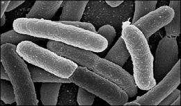


Genomes Of Two Popular Research Strains Of E Coli Sequenced
The genus Salmonella is closely related to Escherichia coli bacteria and is suggested to have diverged from the bacteria (E coli) about 150 million years ago As such, it has adapted and can be found in several niches in the environment Several methods of classification of Salmonella have been suggested so farIn the present study, the synergetic effect and mechanism of ultrasound (US) and slightly acidic electrolyzed water (SAEW) on the inactivation of Escherichia coli ( E coli ) were evaluated The results showed that US combined with SAEW treatment showed higher sanitizing efficacy for reducing E coli than US and SAEW alone treatmentEscherichia coli bacteria on blood agar e coli under a microscope stock pictures, royaltyfree photos & images petri dishes with culture media for sarscov2 diagnostics, test coronavirus covid19, microbiological analysis e coli under a microscope stock pictures, royaltyfree photos & images
E coli under the microscope Escherichia coli (E coli) is a bacterium commonly found in various ecosystems like land and water Most of the strains of E coli are harmless, but some strains are known to cause diarrhea and even UTIs E coli is commonly studied as they are considered as a standard for the study of different bacteriaE coli is a chemoheterotroph whose chemically defined medium must include a source of carbon and energy E coli is the most widely studied prokaryotic model organism, and an important species in the fields of biotechnology and microbiology, where it has served as the host organism for the majority of work with recombinant DNA Under favorable conditions, it takes as little as minutes to reproduceUropathogenic Escherichia coli strains isolated from these patients, as well as the archetypal E coli strain UTI, were found to produce colibactin In a murine model of UTI, the machinery producing colibactin was expressed during the early hours of the infection, when intracellular bacterial communities form
Find the perfect E Coli Under A Microscope stock photos and editorial news pictures from Getty Images Select from premium E Coli Under A Microscope of the highest qualityE coli is a facultative anaerobe 2 Its optimum growth temperature is 37°C and ranges from 10°C to 40°C E coli on Nutrient Agar (NA) 1 They appear large, circular, low convex, grayish, white, moist, smooth, and opaque 2 They are of 2 forms Smooth (S) form and Rough (R) form 3 Smooth forms are emulsifiable in saline 4E Coli E Coli under the microscope at 400x E Coli (Escherichia Coli) is a gramnegative, rodshaped bacterium Most E Coli strains are harmless, but some serotypes can cause food poisoning in their hosts The harmless strains are part of the normal flora of the gut Learn more about E Coli here Helicobacter Pylori
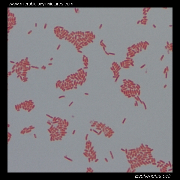


E Coli Gram Stain And Cell Morphology E Coli Micrograph Appearance Under The Microscope E Coli Cell Morphology E Coli Microscopic Picture
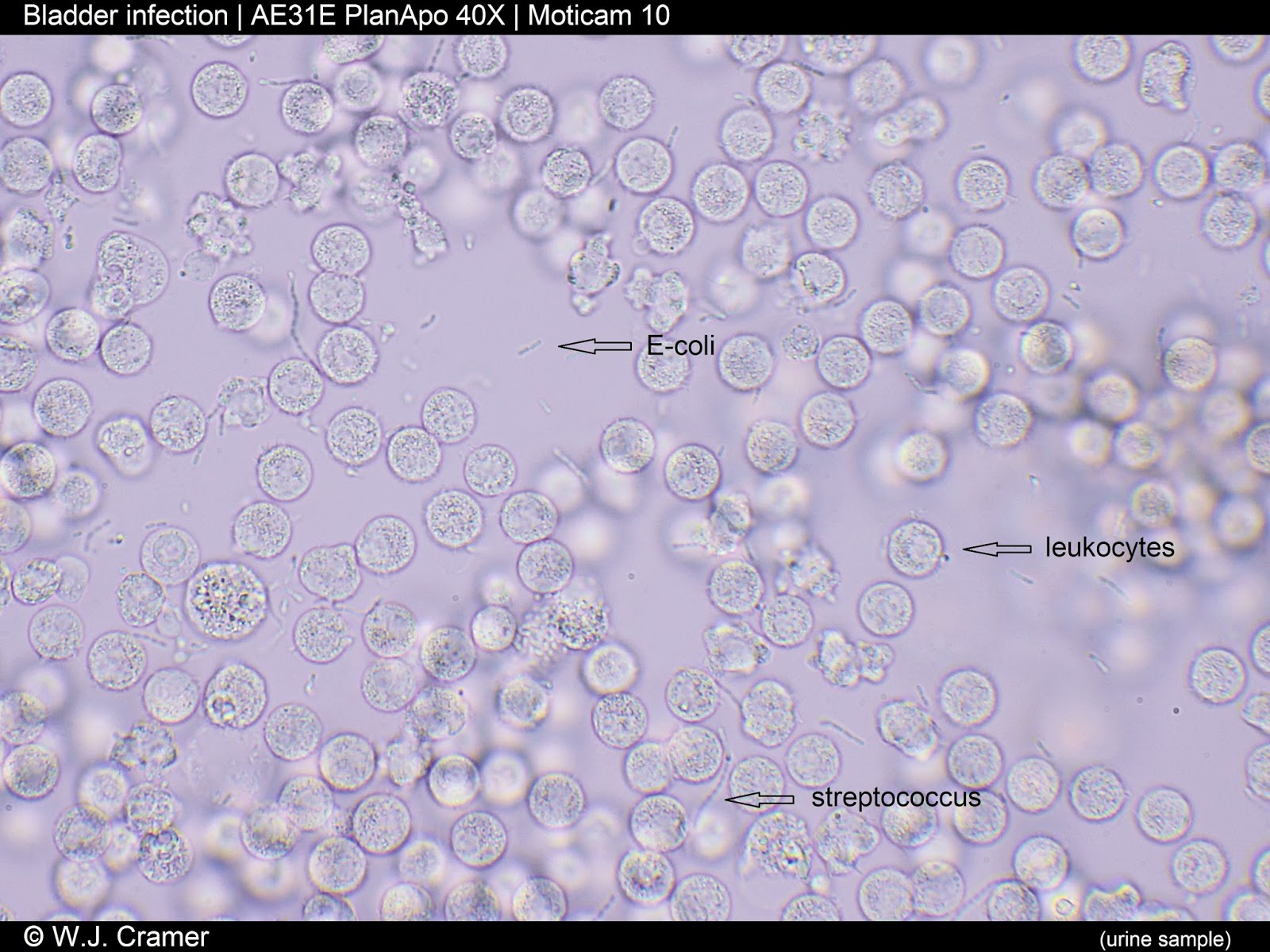


Motic Europe Blog Escherichia Coli Are Usually Blamed
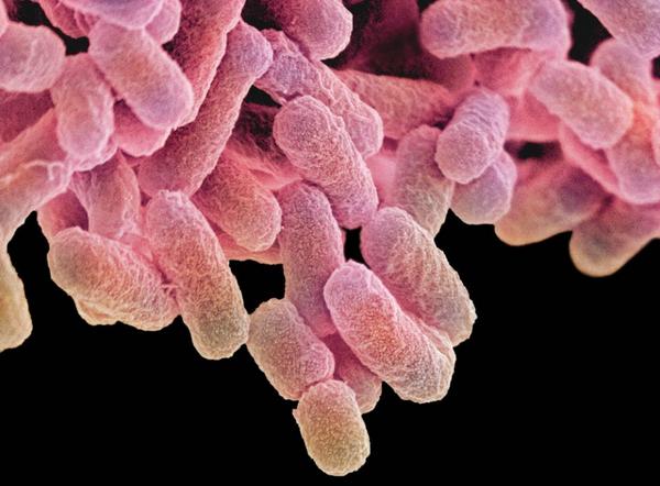
To observe E coli with any detail, you will need to use the 100X lens, which is also known as an oil immersion lens This is the longest, most powerful and most expensive lens on the microscope, requiring extra care when using it As the name implies, the 100X lens is immersed in a drop of oil on the slideE coli, sem e coli microscope stock pictures, royaltyfree photos & images bacterium closeup e coli microscope stock pictures, royaltyfree photos & images Escherichia Coli, Tem, Bacterial Pili, Filamentous Extensions Protruding From The Cell, Which Play An Important Role In Bacterial Adhesion To MucosalSUMMARY The shape of Escherichia coli is strikingly simple compared to those of higher eukaryotes In fact, the end result of E coli morphogenesis is a cylindrical tube with hemispherical caps It is argued that physical principles affect biological forms In this view, genes code for products that contribute to the production of suitable structures for physical factors to act upon


Biol 230 Lab Manual Gram Stain Of E Coli


Figure 3 From Pathologicalstudy On Colibacillosis In Chickens And Detection Of Escherichia Coli By Pcr Semantic Scholar
The Best E Coli Microscope of 21 – Reviewed and Top Rated After hours researching and comparing all models on the market, we find out the Best E Coli Microscope of 21 Check our ranking below 2,9 Reviews ScannedMORPHOLOGY OF ESCHERICHIA COLI (E COLI) Shape – Escherichia coli is a straight, rod shape (bacillus) bacterium Size – The size of Escherichia coli is about 1–3 µm × 04–07 µm (micrometer) Arrangement Of Cells – Escherichia coli is arranged singly or in pairs Motility – Escherichia coli is a motile bacterium Some strains of E coli are nonmotileOn Endo agar it looks like lactose negative)All four strains are mannitol positive (best seen in fig D), cellobiose negative (strains A, B)
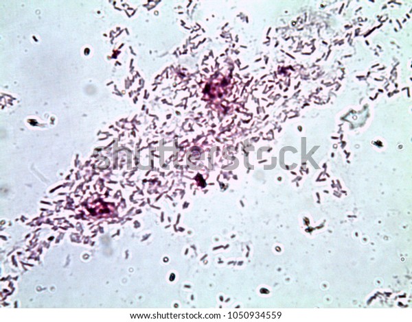


Escherichia Coli Gram Stain Compound Microscope Stock Photo Edit Now


High Precision Characterization Of Individual E Coli Cell Morphology By Scanning Flow Cytometry Konokhova 13 Cytometry Part A Wiley Online Library
The genus Salmonella is closely related to Escherichia coli bacteria and is suggested to have diverged from the bacteria (E coli) about 150 million years ago As such, it has adapted and can be found in several niches in the environment Several methods of classification of Salmonella have been suggested so farPrepared microscope slide of Escherichia coli, bacilli, smear Description This slide of a smear of E coli is best viewed under a microscope with an oilimmersion objective Qty 1 slideLivecell imaging of E coli biofuel synthesis by spectroscopic stimulated Raman scattering microscopy Haonan Lin1,3*(hnlin@buedu), Nathan ue1, Wilson Wong1,4, JiXin Cheng1,2,3 and Mary J Dunlop1,4 1Department of Biomedical Engineering, 2Department of Electrical & Computer Engineering, 3Photonics Center, 4Biological Design Center, Boston University, Boston,
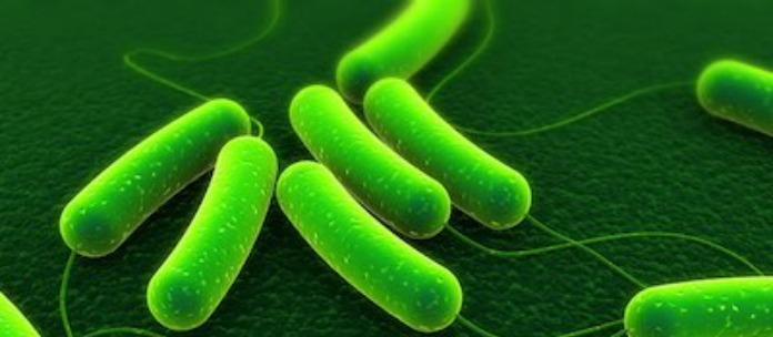


Monroe Montessori School In Wa Closed For Possible E Coli


Escherichia Coli Wikipedia
Escherichia is a gramnegative bacterium, which under the microscope is shaped like a rod with a small tail It is widely distributed in nature (Brooker 08) Escherichia coli (E coli) is part of the normal intestinal flora Some strains are pathogenic and can cause gastroenteritis, UTI, meningitis, and wound infectionsE coli can spread to the urinary tract in a variety of ways Common ways include Your urine will then be examined under a microscope for the presence of bacteria Urine cultureEscherichia is a gramnegative bacterium, which under the microscope is shaped like a rod with a small tail It is widely distributed in nature (Brooker 08) Escherichia coli (E coli) is part of the normal intestinal flora Some strains are pathogenic and can cause gastroenteritis, UTI, meningitis, and wound infections


Q Tbn And9gcs3nlev0tx1pfsddwm6y9ajujk4lxjzmg7ksdxctgnipv0l50c Usqp Cau


Urinary Tract Infections How They Re Prevented Diagnosed And Treated Abc News
E coli under the microscope Escherichia coli (E coli) is a bacterium commonly found in various ecosystems like land and water Most of the strains of E coli are harmless, but some strains are known to cause diarrhea and even UTIs E coli is commonly studied as they are considered as a standard for the study of different bacteriaUropathogenic Escherichia coli strains isolated from these patients, as well as the archetypal E coli strain UTI, were found to produce colibactin In a murine model of UTI, the machinery producing colibactin was expressed during the early hours of the infection, when intracellular bacterial communities formExamples of Bacteria Under the Microscope Escherichia coli Escherichia coli (Ecoli) is a common gramnegative bacterial species that is often one of the first ones to be observed by students Most strains of Ecoli are harmless to humans, but some are pathogens and are responsible for gastrointestinal infections They are a bacillus shaped


Q Tbn And9gctr6jrbwwdcm0wkcm0wnyk6hna6rhc Qg2yjsegfdg1 Pej D2f Usqp Cau
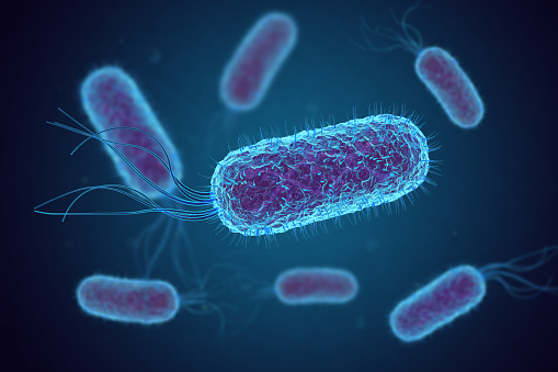


Escherichia Coli Bacteria Under Microscope Stock Photo Download Image Now Istock
Light microscope footage of Escherichia coli bacteria moving around E coli is a Gramnegative bacterium found in the intestines of warmblooded animals It is a normal member of the gut flora, but some strains of E coli can cause food poisoning The bacterial cells are around 2x05 micrometres in size This is the K12 strainLivecell imaging of E coli biofuel synthesis by spectroscopic stimulated Raman scattering microscopy Haonan Lin1,3*(hnlin@buedu), Nathan ue1, Wilson Wong1,4, JiXin Cheng1,2,3 and Mary J Dunlop1,4 1Department of Biomedical Engineering, 2Department of Electrical & Computer Engineering, 3Photonics Center, 4Biological Design Center, Boston University, Boston,In the present study, the synergetic effect and mechanism of ultrasound (US) and slightly acidic electrolyzed water (SAEW) on the inactivation of Escherichia coli ( E coli ) were evaluated The results showed that US combined with SAEW treatment showed higher sanitizing efficacy for reducing E coli than US and SAEW alone treatment


Introductory Chapter The Versatile Escherichia Coli Intechopen


Escherichia Coli Slide W M Science Lab Microbiology Supplies Amazon Com Industrial Scientific
The Best E Coli Microscope of 21 – Reviewed and Top Rated After hours researching and comparing all models on the market, we find out the Best E Coli Microscope of 21 Check our ranking below 2,9 Reviews ScannedEscherichia coli is a bacterium that naturally lives in the intestines of healthy animals and humans, however, there are strains of E coli, specifically O157H7, that cause food poisoning Feces can contaminate food with E coli through runoff and improper sterilization Water, raw vegetables, undercooked ground beef, and unpasteurized milkEscherichia coli Four different strains of Escherichia coli on Endo agar with biochemical slope Glucose fermentation with gas production, urea and H 2 S negative, lactose positive (with exception of strain D "late lactose fermenter";


Bright Field Microscopy Of E Coli Strains Grown To Log Phase In Lb Download Scientific Diagram


27 Earth Bacteria E Coli
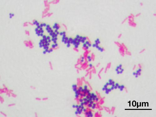
Study E coli, the intestinal bacteria commonly used in labs, with this live cultureE coli is a gramnegative faculative anaerobic bacteria This safe K12 strain of E coli is commonly found in your intestine and is not the kind that causes food poisoning Each culture comes in a test tube on agar media These slant cultures are made with 5ml of agar that is rotated diagonally to provideCocci in grapelike clusters (Saureus) and bacilli(Ecoli) Clinical significance of Saureus Frequently found as part of the normal skin flora on the skin and nasal passages;E coli, sem e coli microscope stock pictures, royaltyfree photos & images bacterium closeup e coli microscope stock pictures, royaltyfree photos & images Escherichia Coli, Tem, Bacterial Pili, Filamentous Extensions Protruding From The Cell, Which Play An Important Role In Bacterial Adhesion To Mucosal


Amazing Electron Microscope Images Page 2 Listoid Microscopy Electron Microscope Electron Microscope Images
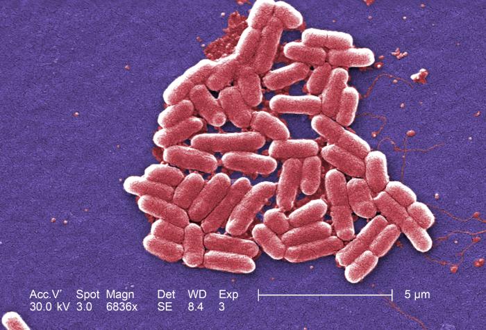


Details Public Health Image Library Phil
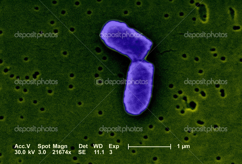
In the present study, the synergetic effect and mechanism of ultrasound (US) and slightly acidic electrolyzed water (SAEW) on the inactivation of Escherichia coli ( E coli ) were evaluated The results showed that US combined with SAEW treatment showed higher sanitizing efficacy for reducing E coli than US and SAEW alone treatmentAllow students to explore the morphology of 1 of the most wellknown, Gramnegative bacteria, E coli This rodshaped, facultative anaerobe is normally present in the intestines of warmblooded animals However, it can be pathogenic This slide works well when discussing harmful and helpful bacteriaUnder a high magnification of 66X, this digitallycolorized, scanning electron microscopic (SEM) image depicted a growing cluster of Gramnegative, rodshaped, Escherichia coli bacteria, of the strain O157H7, which is a pathogenic strain of E coli


Small Things Considered E Coli Cells Face Facs And Get Back Into Shape


Escherichia Coli And Bacillus Sp Microscopy
To observe E coli with any detail, you will need to use the 100X lens, which is also known as an oil immersion lens This is the longest, most powerful and most expensive lens on the microscope, requiring extra care when using it As the name implies, the 100X lens is immersed in a drop of oil on the slideA whole mount of E coli bacteria on a prepared microscope slide (Not recommended for demonstrating typical bacillus morphology) Login or Register • Live Chat (offline) My Account Login or register now to maximize your savings and access profile information, order history, tracking, shopping lists, and moreLivecell imaging of E coli biofuel synthesis by spectroscopic stimulated Raman scattering microscopy Haonan Lin1,3*(hnlin@buedu), Nathan ue1, Wilson Wong1,4, JiXin Cheng1,2,3 and Mary J Dunlop1,4 1Department of Biomedical Engineering, 2Department of Electrical & Computer Engineering, 3Photonics Center, 4Biological Design Center, Boston University, Boston,


Science Source Stock Photos Video E Coli Bacteria Light Microscopy


Gram Negative Pink Colored Small Rod Shape E Coli Under Light Download Scientific Diagram
On Endo agar it looks like lactose negative)All four strains are mannitol positive (best seen in fig D), cellobiose negative (strains A, B)Escherichia coli (abbreviated as E coli) are bacteria found in the environment, foods, and intestines of people and animalsE coli are a large and diverse group of bacteria Although most strains of E coli are harmless, others can make you sick Some kinds of E coli can cause diarrhea, while others cause urinary tract infections, respiratory illness and pneumonia, and other illnessesExamples of Bacteria Under the Microscope Escherichia coli Escherichia coli (Ecoli) is a common gramnegative bacterial species that is often one of the first ones to be observed by students Most strains of Ecoli are harmless to humans, but some are pathogens and are responsible for gastrointestinal infections They are a bacillus shaped bacteria that has a very fast growth (they can double every minutes), which is one of the main reasons they are used in research


Untitled Document


E Coli Growth
E coli is a Gramnegative rodshaped bacteria When Gram stained, the organism looks pink or red Here are a couple of pictures of a Gram stain of E coli that I did under the 100X objective lens on a standard light microscope You can of courseE Coli Microscopy To determine whether a strain (s) is present in a sample, it's necessary to stain the sample Here, Gram stain is used as it helps distinguish between the gram positive and gram negative bacteria in a sample Being a differential stain, Gram stain is more complex compared to more simple stains like methylene blueMost of the bacteria range from 022 µm in diameter The length can range from 110 µm for filamentous or rodshaped bacteria The most wellknown bacteria E coli, their average size is ~15 µm in diameter and 26 µm in length In this figure The size comparison between our hair (~ 60 µm) and E coli (~1 µm) Notice how small the
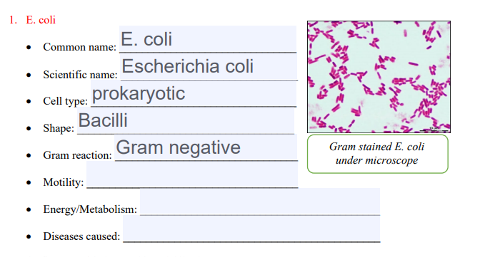


Solved 1 E Coli E Coli Common Name Escherichia Coli S Chegg Com


How E Coli Cells Work In The Human Gut Uconn Today
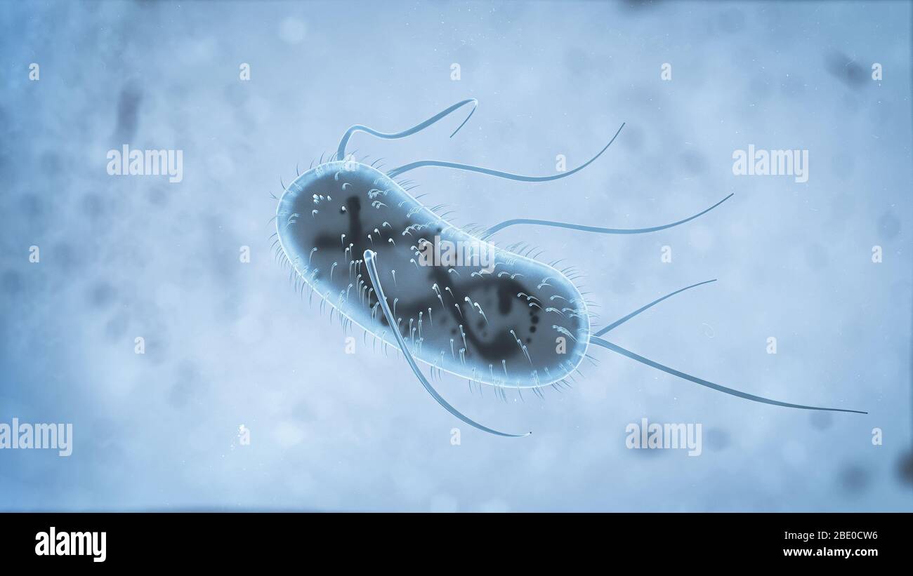
Escherichia coli Four different strains of Escherichia coli on Endo agar with biochemical slope Glucose fermentation with gas production, urea and H 2 S negative, lactose positive (with exception of strain D "late lactose fermenter";Ecoli is a procaryotic microorganism, typically about 2 to 4 µm long, rodshaped bacterium (bacillus) with rounded ends Cells can be shorter than 2 µm or can form long filaments Ecoli is usually motile in liquid or semisolid environment with peritrichous flagella (about 6 per cell) and its surface is covered with fimbriae TheseIt is estimated that % of the human population are longterm carriers of S aureus


Niah Niah Pathogenic Organisms Observed By Electron Microscope Escherichia Coli


Microscopic Studies Of Various Organisms
Uropathogenic Escherichia coli strains isolated from these patients, as well as the archetypal E coli strain UTI, were found to produce colibactin In a murine model of UTI, the machinery producing colibactin was expressed during the early hours of the infection, when intracellular bacterial communities form


Direct E Coli Cell Count At Od600 Tip Biosystems


Escherichia Coli Colony Morphology And Microscopic Appearance Basic Characteristic And Tests For Identification Of E Coli Bacteria Images Of Escherichia Coli Antibiotic Treatment Of E Coli Infections


An Undated File Picture Taken With Electronic Microscope Shows Ehec Bacteria Enterohaemorrhagic Escherichia Coli In Helmholtz Centre For Infection Research In Brunswick Young Scientists Journal


Bayer Pharma Microscope Monday Magnification Of E Coli Bacteria Http T Co U9kwhjsm1g


E Coli Bacteria Light Microscopy Stock Video Clip K004 9042 Science Photo Library


Pin On Microbiology


Escherichia Coli E Coli National Geographic Society


E Coli Escherichia Coli A Gram Negative Bacterium Causing Gastrointestinal Infection


Pictures E Coli Under A Microscope Escherichia Coli Under The Microscope Stock Photo C Imagepointfr


3d Escherichia Coli E Coli Cells Or Bacteria Under Microscope 3d Illustration Stock Photo Alamy
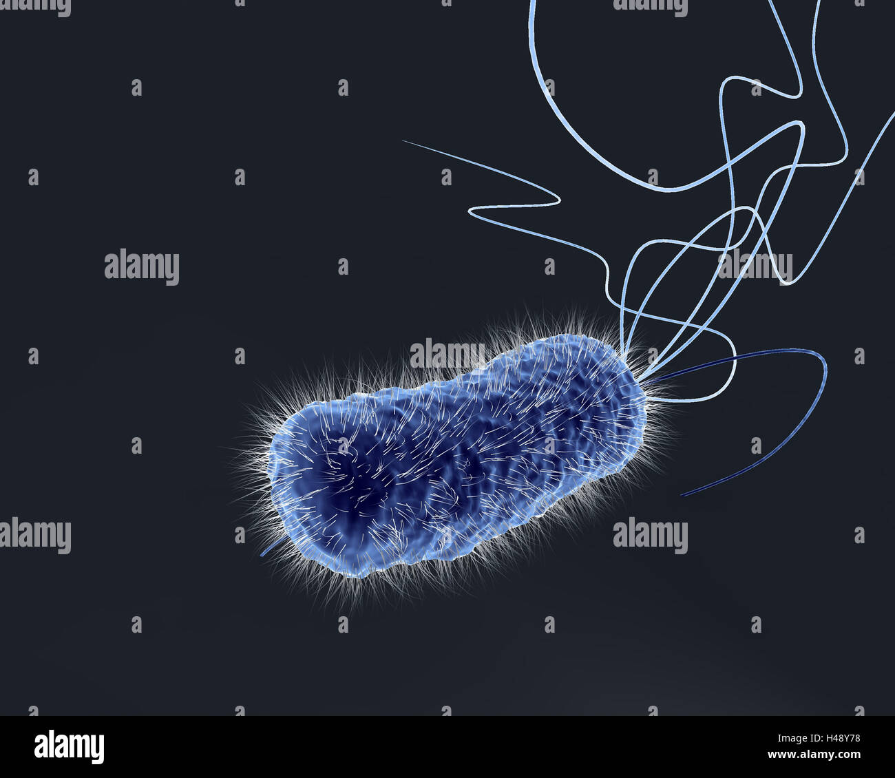


Bowel Bacterium E Coli Computer Graphics Microscope Recording Stock Photo Alamy


Escherichia Coli Wikipedia


What Does An E Coli Bacteria Look Like Under A Microscope Quora


E Coli Bacteria Preventing E Coli Infections


Cytoplasmic Ph Response To Acid Stress In Individual Cells Of Escherichia Coli And Bacillus Subtilis Observed By Fluorescence Ratio Imaging Microscopy Applied And Environmental Microbiology


Introductory Chapter The Versatile Escherichia Coli Intechopen


Staphylococcus Aureus And Ecoli Under Microscope Microscopy Of Gram Positive Cocci And Gram Negative Bacilli Morphology And Microscopic Appearance Of Staphylococcus Aureus And E Coli S Aureus Gram Stain And Colony Morphology On Agar Clinical


Niah Niah Pathogenic Organisms Observed By Electron Microscope Escherichia Coli


Escherichia Coli Light Microscopy


Morphology Of E Coli Cells Under Microscope At 100 Magnification Download Scientific Diagram


Optical Microscope Images Of E Coli Cells Following Gram Staining A Download Scientific Diagram


Part a K Experience Parts Igem Org
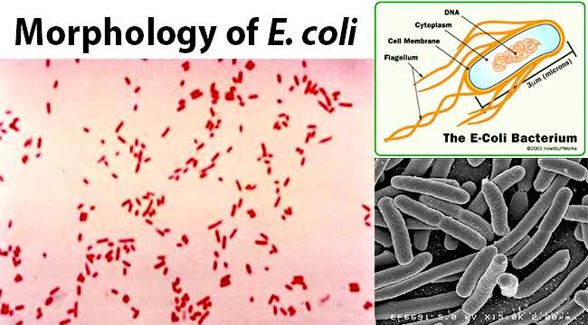


Escherichia Coli E Coli An Overview Microbe Notes
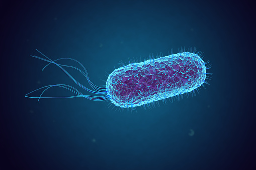


Escherichia Coli Bacteria Under Microscope Stock Photo Download Image Now Istock


E Coli Bacteria Abc News Australian Broadcasting Corporation


Division Of E Coli Bacteria Under The Microscope Video Eurekalert Science News


10pk Escherichia Coli Smear Gram Stain Prepared Microscope Slides 75 X 25mm Classroom Pack 10 Slides In Storage Case Biology Microscopy Eisco Labs Amazon Com Industrial Scientific


Optical Microscope Images Of E Coli Cells Following Gram Staining A Download Scientific Diagram
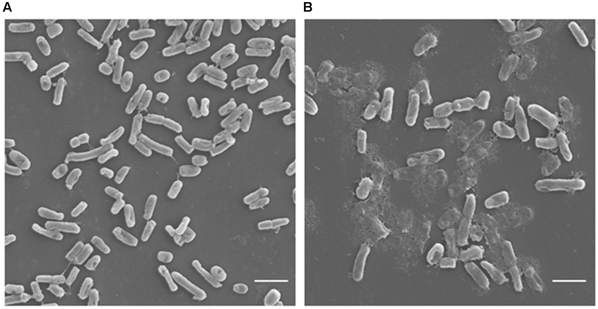


Frontiers Antimicrobial Potential Of Carvacrol Against Uropathogenic Escherichia Coli Via Membrane Disruption Depolarization And Reactive Oxygen Species Generation Microbiology
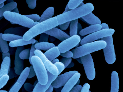


Here S Why Drug Resistant Bacteria Could Spread Globally Time


Bacteria Under The Microscope E Coli And S Aureus Youtube


Transmission Electron Microscopic Micrographs Of E Coli Treated With Peptides


Popular Ballard Restaurant Closed Due To E Coli Outbreak Komo


Gram Stain


E Coli Bacteria Under Microscope Page 1 Line 17qq Com


E Coli Outbreaks In California
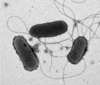


Know Your Enemy Escherichia Coli E Coli Global Food Safety Resource


Eiscoprepared Microscope Slide Escherichia Coli Smear Gram Stain Microbiology Fisher Scientific
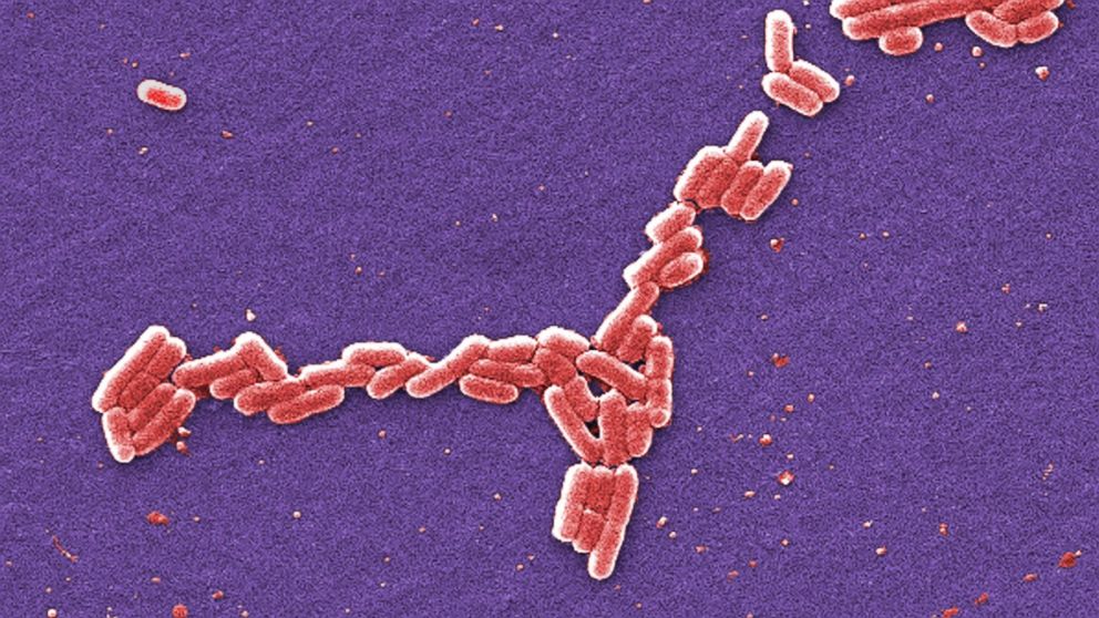


City In Oklahoma Ordered To Boil Water After Possible E Coli Contamination Abc News


Q Tbn And9gctagaczd1acqz3j1t619lvnmx5jx49xxn67stfkqatx5oqst97i Usqp Cau


Lab 1


Self Organization And Cell Morphogenesis Lab Nucleoid


Closer To The End Of Intestinal Infections By Escherichia Coli


Cell Division Of E Coli With Continuous Media Flow Youtube
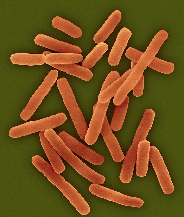


E Coli 0157 H7 Photograph By Dennis Kunkel Microscopy Science Photo Library


Q Tbn And9gcswouuht13c4cxzkdgvwicdnmwhgkfjwlh40a Eerzzlpwjtmyt Usqp Cau


Microbial Contamination From Human And Agricultural Waste Discharges Dossier Environnement Littoral Ifremer


Http Coltonanderson1 Weebly Com Uploads 2 4 3 0 Manual Pdf
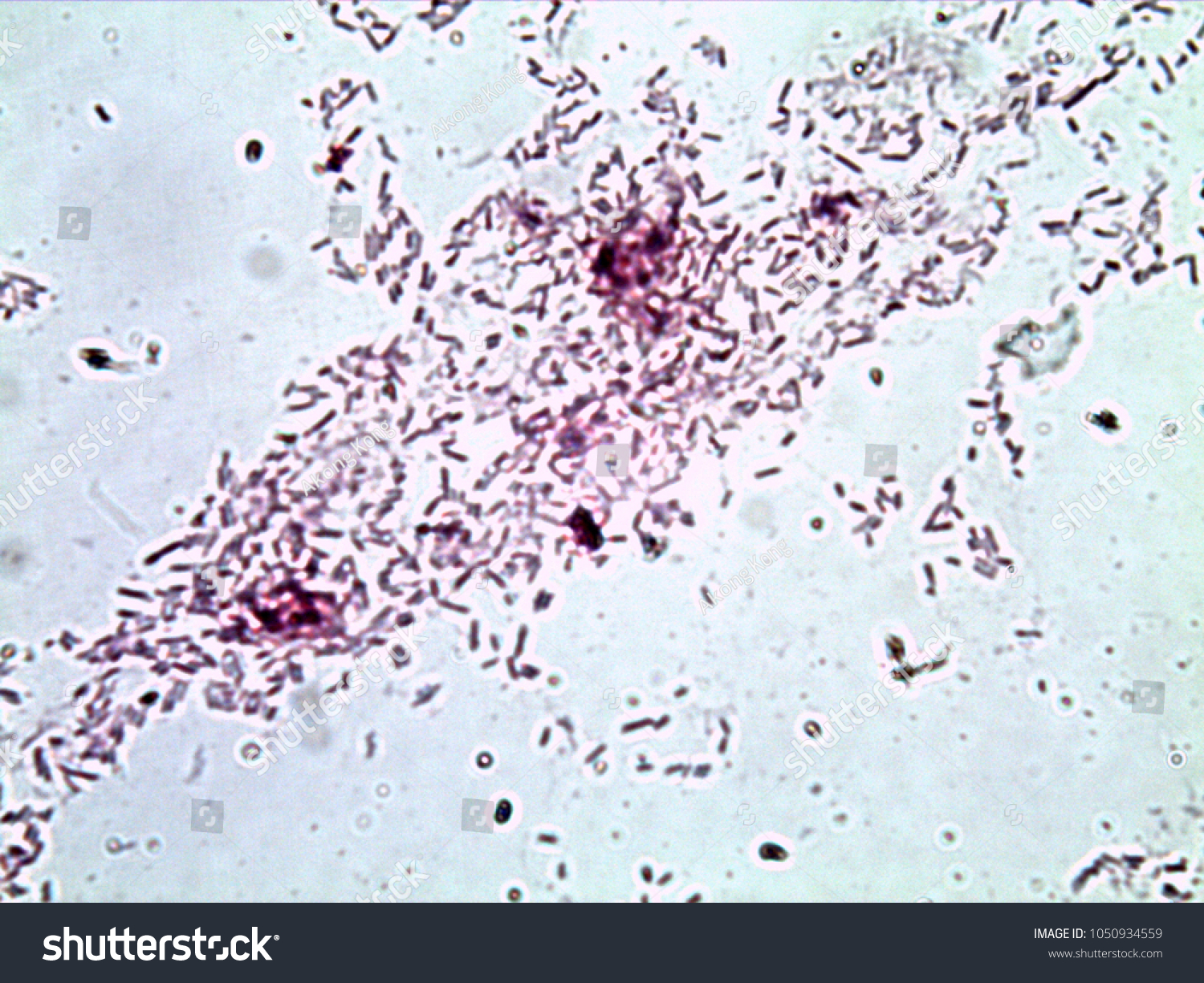


Escherichia Coli Gram Stain Compound Microscope Stock Photo Edit Now


Insight Into Why E Coli Sickness Levels Vary


Image Of The Month E Coli Bacteria Growing On Mini Guts
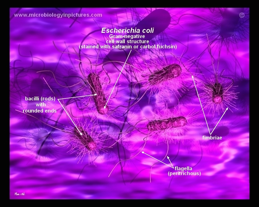


How E Coli Bacteria Look Like
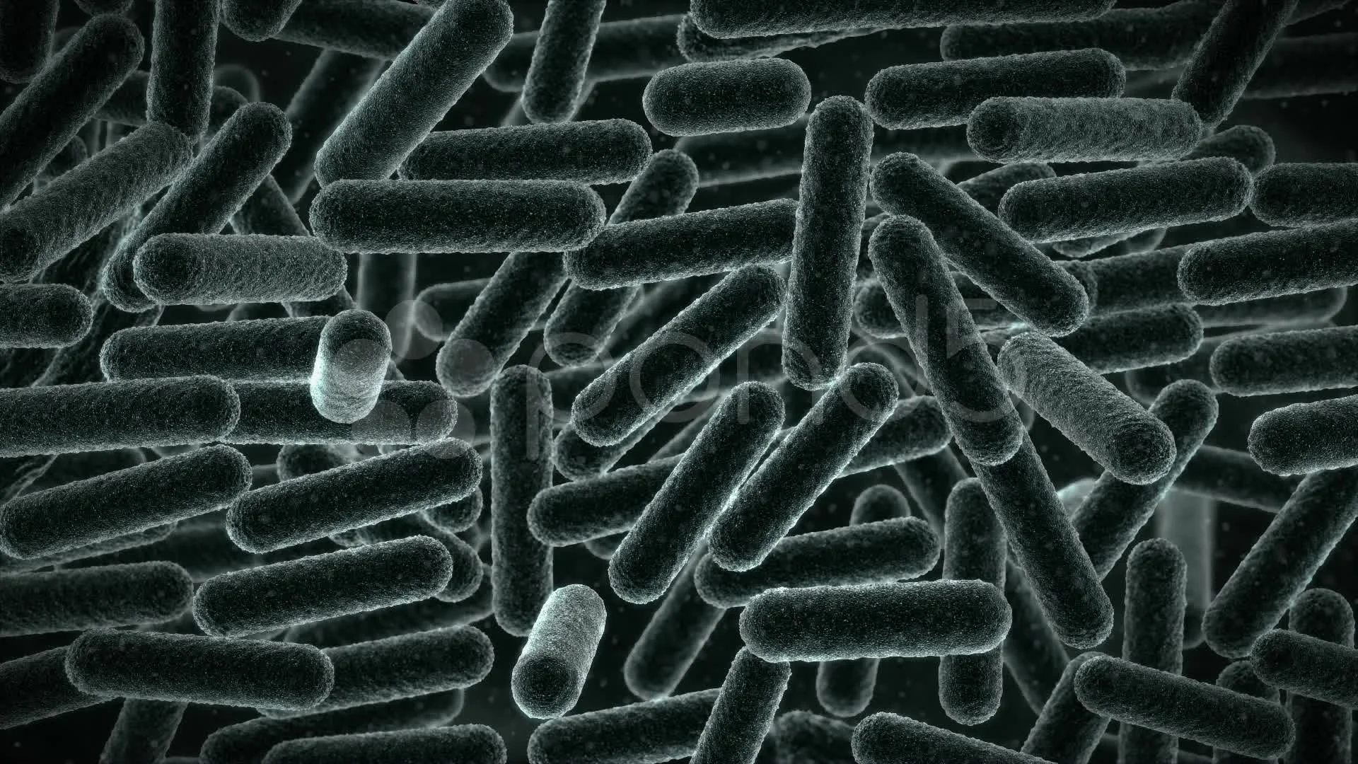


E Coli Virus Electron Microscope View Stock Video Pond5


Aph162 Report 1


Genomes Of Two Popular Research Strains Of E Coli Sequenced


Microscope Imaging Of Methylene Blue Stained E Coli Cells Harbouring Download Scientific Diagram


Escherichia Coli Bacteria E Coli Stock Footage Video 100 Royalty Free Shutterstock
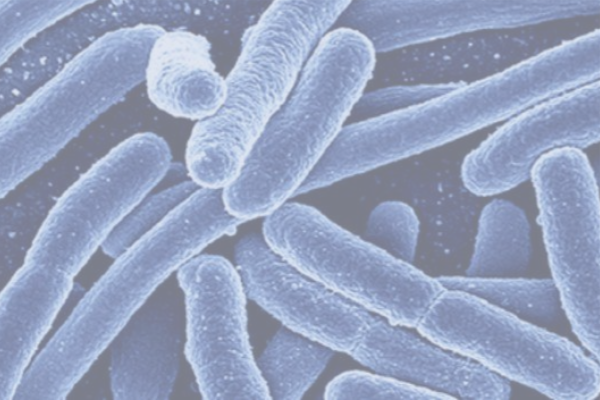


Understanding E Coli In A Food Processing Context Fda Reader
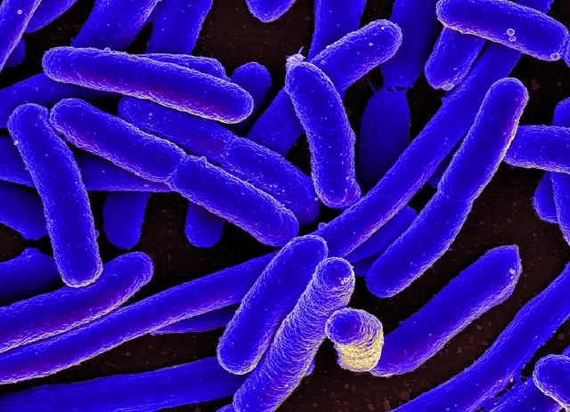


E Coli Under The Microscope Types Techniques Gram Stain Hanging Drop Method


Markus Covert How To Build A Computer Model Of A Cell Stanford School Of Engineering


Pathogenic E Coli


Pathogenic E Coli


E Coli Bacteria Microscopic Organism Indicates Fecal Contamination
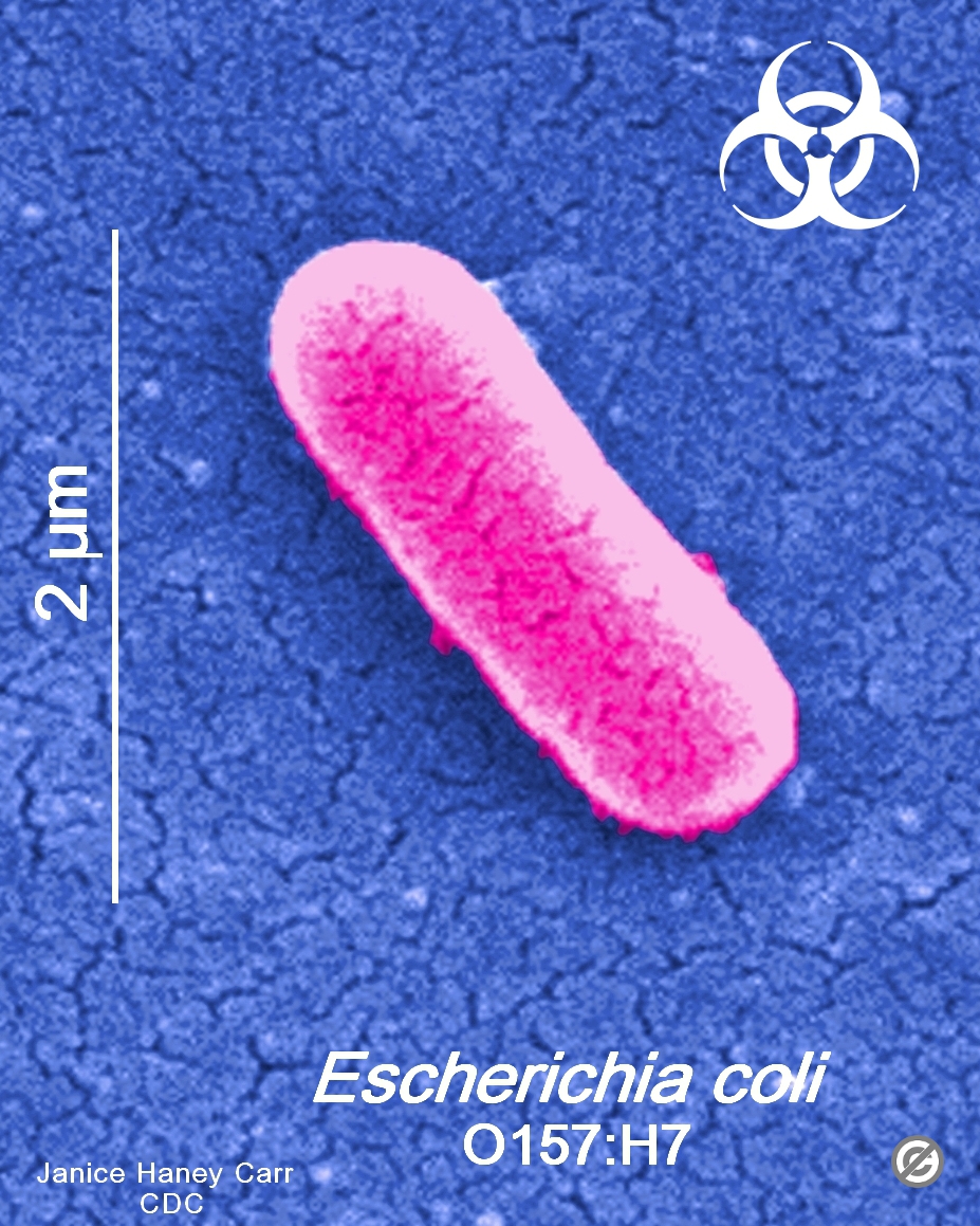


Usa Minnesota Firm Recalls Ground Beef Products Due To Possible E Coli O157 H7 Contamination Food Law Latest
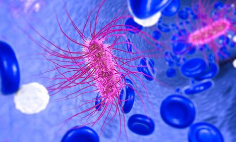


E Coli And Foodborne Illness Fda
.jpg)


Escherichia Coli 400x Escherichia Coli 400x Manufacturers Escherichia Coli 400x Suppliers Escherichia Coli 400x Exporters Escherichia Coli 400x In India


E Coli Png Images Pngegg
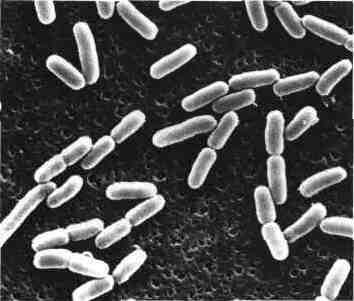


Escherichia Coli Medical Daily News Health News
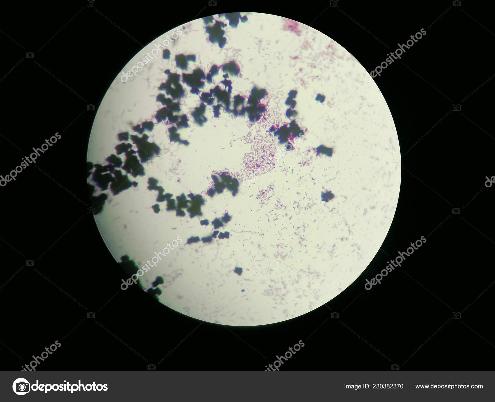


Image Of Escherichia Coli Obtained Through A Light Microscope Stock Photo Image By C Pike 28


Biochip Collaborative University Of Maryland Bentley Group


Production Of Insoluble Starch Like Granules In Escherichia Coli By Modification Of The Glycogen Synthesis Pathway Biorxiv
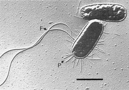


Escherichia Coli 07 Igem Org
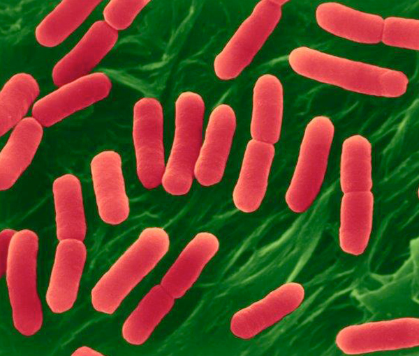


E Coli O157 H7 Escherichia Coli Expert Witness And Epidemiology Services
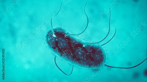


3d Escherichia Coli E Coli Cells Or Bacteria Under Microscope 3d Illustration Buy This Stock Illustration And Explore Similar Illustrations At Adobe Stock Adobe Stock


Slide Escherichia Coli


Escherichia Coli Bacteria Sem Photograph By Niaidcdc


コメント
コメントを投稿